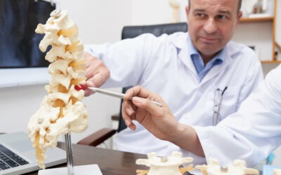
Although the spine looks like a simple framework of 33 stacked vertebrae, it is the most complex structure of the human body after the brain. What’s even more amazing is that the human spine has its own memory
What looks like a simple framework of 33 stacked vertebrae, is actually held in place by over 23 intervertebral discs and 220 special ligaments. Hence spine problems don’t essentially mean bone problems. It could also mean a ligament or a disc issue. Which also makes diagnosing spine problems grueling.
But the healthcare industry, being at the forefront of technology adoption, has come a long way in diagnosing spine problems and improving spine care, all of which is summarized into a single term – Spine Radiology.
This article brings to you a consolidated overview of the advancements in spine radiology.
Table of contents
- What is spine radiology?
- Imaging modalities for spine problems
- Common spine problems
- Challenges in diagnosing spine problems
- Non-surgical treatment options for spine problems
- Surgical treatment options for spine problems
- Quantitative image analysis for spine imaging
- The need for objective reports in spine radiology
- AI and its role in spine care
- Final thoughts
Suggested read: What Are Top Surgeons Looking for in MRI Lumbar Spine Reports?
1. What is spine radiology?
Spine radiology is an umbrella term that refers to various aspects of spine care. Right from using radiography to exploring the various regions of the spine (say cervical, thoracic, lumbar, and sacral regions) to diagnosing and reporting the findings – all activities carried out to study the spine comes under spine radiology.
2. Imaging modalities for spine problems
Back pain is a challenging situation not just for the common man, but also for highly-skilled healthcare professionals, particularly radiologists as it is not easy to identify spine problems at an early stage.
The first step in diagnosing spine problems is examining the person’s gait, ability to sit, stand, and lift the leg. But suspicions are confirmed through medical images like x-rays, CT scans, and MRIs.
2.1. X-rays
X-rays are the quickest, pain-free, and noninvasive imaging procedures that are most commonly used in spine radiology. X-rays are used to check for spine alignment issues, changes in the spine due to osteoarthritis, fractured vertebrae, etc. However, physicians might order a different imaging study if they suspect a disc or muscle, or nerve tissue in the spine.
2.2. Stress x-rays
Stress x-rays, which are taken after applying required stress to a particular area, are used to detect ligament damage, evaluate injuries, and determine their severity. Stress x-rays also play a major role in identifying excessive motion, evaluating the degree of soft tissue damage, and determining conditions like arthritis, disc degeneration, spondylolisthesis, tumors, etc. They also aid in clinically correlating areas of spinal instability.
2.3. CT scan
CT scan is a collection of multiple x-rays produced at different angles to create a complete 3-D view of the soft tissues and bones. They are primarily used to diagnose back injuries associated with soft tissues and produce high-clarity images of organs, muscles, and ligaments as compared to a conventional x-ray.
CT scans are mostly used in emergency cases like accidents and the diagnosis of ruptured disc, disc degeneration, spinal stenosis, tumors, spinal cord infections, etc. CT scans can also prevent/reduce the need for exploratory surgeries.
2.4. MRI
Similar to CT scans, MRI scans also produce highly detailed images of the spine. But the difference is that MRI uses magnetic fields and radio waves to generate the images instead of x-rays. Physicians order MRI during suspicions of disc rupture, infection, tumor, etc, in the spine. Though MRI is a painless and non-invasive spine imaging procedure, it may not be suitable for people suffering from claustrophobia or allergies.
2.5. X-rays vs. MRI vs. CT scan: Which is the best?
All the imaging modalities come with their benefits and risks.
Both x-rays and MRIs are painless procedures. X-rays are highly instrumental in identifying bone tissues and are a faster procedure compared to MRI. On the other hand, MRI is a better option for diagnosing soft tissue problems as it eliminates the need for exposing patients to ionizing radiation to capture images.
Similarly, CT scans are also a quicker procedure compared to MRIs. But CT scans are generally used for diagnosing severe injuries that require immediate attention. CT scans are also helpful in spotting tumors. Though CT scans are better than MRI in capturing bone images, patients are exposed to a small dose of radiation.
3. Common spine problems
Spine problems have become ubiquitous and age-agnostic all over the world. Research states that 80% of the globe’s population will experience back pain at some point in their lives. Besides, spine problems have more adverse social and economic effects as compared to life-threatening diseases like cancer. Nearly 264 million man-days are lost annually on account of back pain.
It’s not just the complex structure of the spine that makes it hard to narrow down the actual problem. Spine conditions often co-occur. For instance, a person suffering from a slipped disc can also have spinal stenosis. In most cases, only one condition is diagnosed.
Here are some of the common spine problems that the majority of the globe’s population are suffering from:
3.1. Disc degeneration
Disc degeneration is one of the most common spine problems that every individual is likely to experience as they age. According to stats, over 40% of people above the age of 40 have at least one degenerated disc.
But, disc degeneration is asymptomatic until it worsens which makes it increasingly difficult for physicians to identify at an early stage. Also, disc degeneration and multiple sclerosis share the same symptoms, which can mislead the physician during a diagnosis.
Today, advancements in technology are enabling physicians to identify asymptomatic conditions like disc degeneration at an early stage through quantitative imaging analysis.
3.2. Ligament sprains and muscle strains
Ligament sprain and muscle strain are the most common reason for acute back pain and can also create painful spasms in the spine. Ligament sprain and muscle strain occur due to numerous reasons like improper lifting of objects, over-stretching, and picking up bulky objects.
In the case of ligament sprains and muscle strains, diagnostic tests are not necessary unless the pain continues for over 6 weeks and has not improved as expected after physical therapy. Since ruling out other underlying causes like disc injury is important, physicians order imaging tests like x-rays and MRIs when symptoms persist for longer than six weeks and physical therapy has not improved the condition.
3.3. Spinal stenosis
Spinal stenosis is a condition that occurs due to the narrowing of the spinal canal. This can put pressure on the nerves passing through the spine, creating pain or numbness. Spinal stenosis, which mostly affects the neck and the lower back, is often the result of osteoarthritis and is prevalent among adults of age 50 and above.
While spinal stenosis can be easily identified through physical exams and imaging tests, the severity of the condition is determined as per the radiologist’s perception. Hence, mild spinal stenosis can be misinterpreted as moderate or vice versa. Such misinterpretations are often the result of subjective analysis or intra-observer variability.
While images can be deceiving, numbers never lie. This is why measuring spine elements (or quantitative analysis) is more effective than subjective analysis and improves the accuracy of diagnosis for both the disease and its severity.
Technologies like deep active learning and machine learning are currently enabling physicians to do a detailed study of images by providing accurate measurements and characterization of spine elements. Even the small elements like neural foramina that can be misread during eyeballing can be avoided with the help of such technologies.
3.4. Skeletal irregularities
Skeletal irregularities refer to the abnormal alignment of the vertebral column. The common types of skeletal irregularities include:
Lordosis – extreme inward curving of the lumbar spine
Kyphosis – forward rounding of the upper back
Scoliosis – sideways curvature of the spine
But such skeletal deformities, particularly scoliosis generally don’t cause back pain until middle age.
AI algorithms can now measure cobb angle and draw stress lines, which makes it easier for the physician to determine the severity of scoliosis.
3.5. Osteoporosis
Osteoporosis is a bone disease that occurs when the bone density decreases, affecting the strength and structure of the bone, ultimately resulting in fracture. Osteoporosis often occurs in the spine and increases the risk of spine fractures. This condition makes the bones so brittle that even bending and coughing can cause fractures.
Nearly 200 million people across the globe suffer from osteoporosis. The US alone accounts for 54 million people with osteoporosis.
Although osteoporosis is a silent disease and is difficult to diagnose at an early stage (as the disease progresses without any pain or symptoms and is found only when the bones fractures), it is a preventable and treatable disease.
Osteoporosis can be diagnosed at an early stage by measuring vertebral height. But since osteoporosis is asymptomatic, physicians don’t suspect it or measure the vertebral height. Even then osteoporosis can be identified at an early stage with the help of AI.
AI algorithms can measure all the spine elements, including vertebral height. By comparing AI’s findings against the anatomical value. physicians can determine osteoporosis before the occurrence of a fracture.
3.6. Traumatic injury
Traumatic injuries such as injured muscles, torn ligaments, ruptured disc, etc., caused by accidents, playing sports, falling down the stairs, etc., can lead to back pain. The lumbar spine is the most prone to traumatic injuries. According to NCBI’s report, 48% of traumatic injuries on the spine are caused due to motor vehicle accidents, 21% by falls, and 14.6% by sports injuries.
Non-traumatic injuries are caused by cancer, inflammation, arthritis, infections, disc degenerations, etc. Whether traumatic or non-traumatic, injuries to the spine affect the nerve fibers passing through the injured area and can impart all or part of the nerves and muscles below the site of injury. Spine injuries often affect:
- Bowel and bladder incontinence
- Heart rate
- Breathing
- Sensation and reflexes
- Muscle movement
- Metabolism
With AI, any type of traumatic injury can be thoroughly studied. Not just that, AI also helps in medical claim scrubbing. Physicians and providers no longer have to fall prey to incorrect claims with AI.
4. Challenges in diagnosing spine problems
As a radiologist or a general physician, how many times did the test results were different from your suspicion? Or how many times has a patient re-visited for back pain within a short time after successfully treating it?
Too many to count?
Research shows that only 10% of people find the root cause of their back pain. It’s not just that the spine is a complicated framework. There are numerous other reasons like the following that make diagnosing spine problems increasingly challenging:
4.1. Pain is a subjective experience
Every person has a different description of pain. What might seem mild or tolerable to one individual might seem extremely painful to another. And a physician’s suspicion is based purely on the individual’s description of back pain.
For instance, a patient might have a herniated disc, but according to the individual, the pain is moderate and tolerable. So the physician might not suspect a herniated disc and suggest non-surgical treatments like exercise and physical therapy for pain relief.
4.2. Back pain can be the result of a combination of problems
Back pain does not stem from one kind of spine problems like spinal stenosis or ruptured disc. It is often the result of a combination of problems, which makes it increasingly difficult to identify the actual cause of pain. Not just physical factors, but even psychological factors like stress, depression, and anxiety can also lead to back pain.
So a patient can have both herniated disc and spinal stenosis at the same time and can be experiencing back pain due to any one of the two. But the physician might not diagnose both conditions. But, as the treatment varies for both conditions, missing out on any one of the conditions can result in carrying out inaccurate treatment and may not help in eliminating the pain source.
Meanwhile, the undiagnosed spine ailment can worsen during inaccurate treatment and might result in appalling outcomes.
4.3. Limited value of diagnostic tests in spine problems
Diagnostic tests have limited value in diagnosing spine problems as there is no single diagnostic test that helps narrow down the actual cause of back pain. Furthermore, certain diagnostic tests such as diagnostic nerve block injections can produce false-negative or false-positive results.
Though there is no single diagnostic test for diagnosing spine problems, it doesn’t mean that diagnostic tests cannot help in diagnosing spine problems. Common spine conditions like lumbar herniated discs that create symptoms of sciatica can be quickly and accurately diagnosed through a couple of clinical tests followed by imaging tests and questionnaires.
5. Non-surgical treatment options for spine problems
Spine problems are undoubtedly complicated. Which often makes people think that spine problems can be better treated only through surgeries and seeking professional help at the initial stage. Most times, individuals seek medical help only when the pain goes intolerable.
But the fact is that only 10% of spine problems require surgery or any form of minimally invasive procedure. 90% of spine problems can be well treated without surgery and with simple lifestyle changes.
Here’s a range of non-surgical options for treating spine problems:
5.1. Exercise
The most economical and easy way to treat back pain is to move more. Research states that exercising for just 30 minutes, 3 to 4 times every week helps in preventing and reducing back pain. One of the simple exercises that most physicians recommend for back pain patients is walking. Besides pain, the other reason why doctors recommend moderate exercises like walking is that it helps in maintaining a healthy weight, which is essential for pain management and maintaining spine health.
5.2. Medications and injections
Anti-inflammatory medications and injections like facet blocks, epidurals, selective nerve root blocks, etc., are used for managing pain for conditions like sciatica and arthritis.
5.3. Physical Therapy (PT)
Physical therapy is one of the most important non-surgical treatment options for treating back and neck pain.
Expert-guided physical therapy helps in strengthening and conditioning spinal muscles, tendons, and ligaments, including the adjacent body structures. However, physical therapy is not an option to postpone spine surgery, but a great way to reduce the pain, particularly when surgery is not helping.
Also, almost all patients who have had spine surgery go for physical therapy to accelerate recovery and regain strength. With the help of AI and its findings, physical therapists and chiropractors can treat spine injuries more safely, avoiding future complications.
5.4. Functional Electrical Stimulation (FES)
Functional Electrical Stimulation(FES) is a method where low-level electric pulses are applied to weakened or paralyzed muscles to restore or improve in people having spine injuries. FES is generally performed by any spine specialist or sports medicine chiropractor.
Functional Electrical Stimulation can also be used to block pain message signals and restore/improve other bodily functions like bladder and bowel control.
5.5. Myofascial release
A myofascial release is a form of physical therapy often used in massage to treat skeletal muscle immobility. While myofascial release can’t heal nerve damage, it can significantly help in reducing pain and regaining movement.
6. Surgical treatment options for spine problems
Surgeries on the spine are done only when necessary or in case of emergency like spinal injuries from motor vehicle accidents.
But spine surgeries don’t always mean cut-open surgeries. There are also minimally invasive procedures that can be used to treat spine conditions. Here’s a brief overview
of the surgical treatment options for spine problems:
6.1 Minimally invasive procedure
Minimally invasive procedures are used to treat spine conditions of severity mild to moderate. In minimally invasive procedures, an endoscope (a tiny lighted tube with a camera on its tip) enters the spine to treat the condition without much damage to the surrounding muscles and tissues. Minimally invasive procedures also help in reducing infection risk and recovery time.
Spine conditions that can be well-treated through minimally invasive procedures:
* Herniated disc (mild to moderate) – removing just the damaged part through an endoscope
* Spinal stenosis (Lumbar region) – extracting bones and tissues that crowd the nerves
* Scoliosis (mild to moderate) – fusing the affected areas of the spine and straightening it through small incisions
* Spinal fractures (mild to moderate) – using small balloons to inflate the vertebral parts to their actual position and using bone cement, a body-safe adhesive, to help with pain caused by fractures
* Spondylolisthesis – using small incisions to fuse the affected areas and keep the vertebrae in their appropriate spaces
* Small spinal column tumors – using small incisions to remove just the tumor and the adjacent affected tissue
6.2 Traditional open surgery
Traditional open surgery is preferred for more complex conditions and patients who have scar tissue from previous spine surgeries or those who require surgical implants like rods and pedicle screws. Spine conditions that require traditional open surgery include:
* severe disc degeneration
* severe spinal fractures
* severe scoliosis
* spinal instability
* spine tumors – large and complex
* spine trauma caused by motor vehicle accidents.
Open surgery is the last option for spine injuries unless it is not a traumatic injury that needs immediate medical attention and where open surgery is the only option.
With the help of AI, most spine conditions like ligament tear, herniated disc, osteoporosis, tumor, etc, can be easily identified at an early stage and effectively treated through minimally invasive procedures, avoiding high-cost spine treatments and longer hospital stays for patients.
7. Quantitative image analysis for spine radiology and spine care
If you are a practicing physician, then you must have come across patients looking for a second opinion and cases where your diagnosis was different from the previous one.
As you know, in the current radiology workflow, analysis and reporting are carried out subjectively. This increases diagnostic errors and inter-observer variability – all of which affect the delivery of spine care at the right time.
An observational study to compare interpretive findings from 10 different MRI centers for one patient’s scan, showed that there wasn’t a single finding that was mentioned across all the reports. Besides, nearly 49 distinctive findings related to a distinct pathology were reported.
Misdiagnosis and inter-observer variability which are highly detrimental to patient care can be significantly reduced, if not eliminated with the help of quantitative imaging analysis.
Quantitative imaging analysis is performed by quantifying the measurable features of medical images and comparing them against anatomically and physiologically relevant metrics to determine diseases and assess their severity.
Quantitative imaging analysis enables comparative analysis of images from different timelines, allowing radiologists and physicians to understand disease evolution and progression.
Using quantitative imaging analysis, radiologists can perform a detailed study of spine images and diagnose spine problems like disc degeneration at an early stage. Quantitative imaging analysis also helps in improving the clinical value of reports for surgeons.
8. The need for objective reports in spine radiology
Reports generated from quantitative imaging analysis are known as objective reports.
Given the current lifestyle, spine problems are likely to become the globe’s next endemic. It is high time that health systems focus on improving spine radiology and thereby spine care. Performing quantitative image analysis and creating objective reports can be the first step toward providing better spine care.
8.1 Improved communication among healthcare professionals
The communication gap is one serious issue that has been hindering health systems from providing optimal patient care for years. Despite actively embracing digitization and integrating technological innovation, healthcare providers haven’t been able to successfully reduce or eliminate communication gaps in their institutions.
One of the key reasons for the increased communication gap in health institutions is subjective analysis and reporting. Reports, which are one of the most basic modes of communication among healthcare professionals, often lack the required clinical value for surgeons and post-operative care. Industry-leading physicians have claimed that surgeons reach out to images directly and don’t refer to radiology reports as they lack the required clinical value
Objective reports give a clear picture of the patient’s condition while increasing the clinical value of reports for physicians and surgeons. Also, objective reports can reduce misinterpretations drastically.
8.2 Choosing the appropriate treatment and surgery
Out of every hundred spinal surgeries, two wrong surgeries are performed due to wrong measurement and characterization
Objective reports can help in choosing the right course of treatment and surgery for spine problems. Consider this – a herniated disc of 2mm or 3mm can be treated through a minimally invasive surgery instead of traditional open surgery. But in subjective reporting, the finding will only be recorded as “Condition – herniated disc, Severity – mild or moderate (depending on the radiologist’s interpretation)”.
If the severity is recorded as mild, then the surgeon would confidently opt for a minimally invasive procedure. But if the severity is recorded as moderate (which is between 3mm – 5mm) surgeons would go for an open surgery thinking that the herniated disc should be of considerable size and cannot be operated through a minimally invasive procedure.
Objective reports prevent incorrect treatments or unnecessary open surgeries that might increase expenditure and hospital stay for patients.
9. AI and its role in spine care
Undoubtedly, quantitative imaging analysis and objective reports will improve spine radiology and spine care. But performing quantitative imaging analysis and generating objective reports for every imaging test is not as easy as it sounds. Particularly not in the current healthcare climate, where the use of imaging tests is increasing at an alarming rate and radiologists’ shortage is growing at a dismal rate.
Research shows that radiologists read 78% more cases today as compared to 2008. Not just that, radiologists are opting for early retirement as a result of burnout. According to statistics, 50% of radiology retirees are under the age of 55, which was just 20% in 2018.
Quantitative imaging analysis is not possible without AI. Using AI’s help, computers can be trained to think and act like radiologists.
With AI not only can health systems perform quantitative imaging analysis and generate objective reports seamlessly, but they also reduce radiologists’ workload and associated burnout by automating repetitive mundane tasks like image sorting, workload distribution, prioritization of cases, etc.
In the case of spine radiology, AI can do more than just sorting and prioritizing cases. AI can automate spine reading by locating and labeling vertebrae, marking morphological points, identifying pathologies and grading their severity, etc.
Suggested read: AI Is Moving from Cancer Diagnosis to Spine Radiology: 5 Ways How AI Will Help Radiologists
Final thoughts
The spine is one of the most sensitive areas of the human body as it accounts for a person’s mobility. Hence it is paramount to make a timely and accurate diagnosis of spine problems so that health systems can provide the optimal patient care at the right time. And the best way to provide the best spine care possible is to switch to quantitative imaging analysis and objective reporting.
80% of reporting time in spine MRIs is spent on tasks that can be automated. Numerous AI algorithms are currently being developed to automate spine reading.
Spindle is one such AI tool built for automating the reading and reporting of spine MRIs. The tool reduces 60% of radiologists’ time spent on spine MRIs by automatically sorting bad quality images, prioritizing critical cases, locating and labeling vertebrae, marking morphological points, grading disc degeneration, detecting nerve root compression, evaluating – lateral recesses, epidural lipomatosis, ligamentum flavum thickening, etc. Spindle can also generate objective reports in just a click.
Want to overcome the challenges in spine reading and reporting? Check this 2-minute video.